Перевести на Переведено сервисом «Яндекс.Перевод»

Technology prolong life

Database
Diet of Longevity (Caloric Restriction)

Authors:

C. M. McCay, R. Walford, R. Weindruch.

History:

In 1930s, C. M. McCay found in his researches that a dietary regimen that reduces calorie intake but maintains micronutrient levels resulted in the increase of the maximum and medium life spans of rats and mice 30–50%. Last years, this technology, due to its simplicity and stable reproducibility, has become one of the leading models in the investigations of fundamental mechanisms of aging and life extension.

Example:

A study of a lifespan was made by the University of Wisconsin in 1989 — 2014. It involved nonhuman primates (rhesus monkeys) and found that caloric restriction primates were only 36.4% as likely to die from age-related causes when compared with control animals, and had only 56.2% the rate of death from any cause.

Description:

Increasing evidence suggests that energy balance is central to both successful ageing and protection from metabolic disorders.
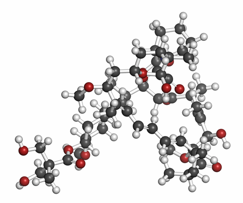
Energy restriction, also known as caloric restriction, is currently the only dietary intervention that is proven to increase longevity and delay the onset of age-related decline in a wide variety of organisms, including Caenorhabditis elegans, Saccharomyces cerevisiae, Drosophila melanogaster, rodents. Nevertheless, in primates CR appears to not affect longevity, but clearly delay age-associated disorders improving health.
Calorie restriction, or caloric restriction (CR), is a dietary regimen that is based on low calorie intake but without malnutrition. «Low» can be defined relative to the subject’s previous intake before intentionally restricting calories, or relative to an average person of similar body type.
Even though there has been research on CR for over 70 years, the mechanism by which CR works is still not well understood. Some explanations include reduced core body temperature, reduced cellular divisions, lower metabolic rates, reduced production of free radicals, reduced DNA damage and hormesis.

Additions and Criticism:

It should be noted that long-term calorie restriction at a level sufficient for slowing the aging process is generally not recommended in children, adolescents, and young adults (under the age of approximately 21), because this type of diet may interfere with natural physical growth and mental development. Pregnant women and women trying to become pregnant are advised not to practice calorie restriction, because it may result in ovulatory dysfunction (infertility), and underweight mothers are more prone to preterm delivery.

Publications:

  • Spindler, Stephen R., Joseph M. Dhahbi, and Patricia L. Mote. «Protein turnover, energy metabolism, aging, and caloric restriction." Advances in cell aging and gerontology 14 (2003): 69–86.
  • Ungvari, Zoltan, et al. «Mechanisms Underlying Caloric Restriction and Lifespan Regulation Implications for Vascular Aging." Circulation research 102.5 (2008): 519–528.
  • Mattison, Julie A., et al. «Impact of caloric restriction on health and survival in rhesus monkeys from the NIA study." Nature (2012).
  • Willcox, Bradley J., et al. «Caloric restriction, the traditional Okinawan diet, and healthy aging." Annals of the New York Academy of Sciences 1114.1 (2007): 434–455.

Geroprotectors (anti-aging drugs)

Authors:

Anisimov v. N., Skulachev v. P., Moskalev A.A.

History:

The ancient philosophers did not want to put up with aging and death. And the ancient alchemists strived to create an elixir of immortality. People were looking for aids to make life longer, or at least to provide healthy aging. At first they used herbs and roots; and with the progress of Molecular Biology and Biotechnology, the scientists tended to fight aging at a molecular level. Therefore, up to date, it were discovered and studied more than 40 chemical geroprotectors.

Example:

Scientists have found that metformin and other biguanides reduce the total mortality rate by more than one third, as well as the mortality from heart attacks and complications from diabetes mellitus; improve the survival rate of cancer patients, and also reduce the risk of breast cancer in patients with type 2 diabetes.

Description:

Geroprotectors (anti-aging drugs) — a common name for a group of substances possessing the ability to increase the lifespan of experimental animals and human.

Up to date, it were discovered and studied more than 40 chemical geroprotectors with declared efficacy. Among them the most famous are resveratrol, rapamycin, protein biosynthesis inhibitors (olivomycin, actinomycin), hormones (growth hormone, thyroid hormones, adrenocortical hormones, reproductive hormones, melatonin), biguanides (metformin, phenformin and others).

Among the mentioned geroprotectors metformindeserves a special attention. The results of studies on mammalian have led to the hypothesis that hyperinsulinemia and hyperglycemia are very important factors both in aging process and in cancer development.

In clinical observations it was found that metformin and other biguanides reduce the total mortality by more than one third, as well as the mortality from heart attacks and complications from diabetes mellitus, improve the survival rate of cancer patients and also reduce the risk of breast cancer in patients with type 2 diabetes. In experiments on laboratory rodents it was discovered a geroprotective effect of antidiabetic medicines, accompanied with decreasing of frequency of spontaneous tumours development. In different models of chemical and radiation carcinogenesis it was found that biguanides are able to slowdown the induced tumours development, and inhibit the growth of many transplanted tumours. 

Additions and Criticism:

Most authors take the view that until now there is no one chemical geroprotector with incontestable proven effect.

Publications:

  • Anisimov, Vladimir N., et al. «If started early in life, metformin treatment increases life span and postpones tumors in female SHR mice." Aging (Albany NY) 3.2 (2011): 148.
  • Morrison, John A., Elizabeth M. Cottingham, and Bruce A. Barton. «Metformin for weight loss in pediatric patients taking psychotropic drugs." American Journal of Psychiatry (2014).
  • Rattan, Ramandeep, et al. «Metformin suppresses ovarian cancer growth and metastasis with enhancement of cisplatin cytotoxicity in vivo." Neoplasia 13.5 (2011): 483-IN28.

Gene Therapy

Authors:

Development of this technology is links with such names as S. Rogers, T. Friedmann and R. Roblin.

History:

The concept of gene therapy arose during the 1960s and 1970s. By January 2014, some 2,000 clinical trials had been conducted or approved.

Example:

The first successful case of gene therapy occurred in the 1990s on a young girl named Ashanti Desilva, a victim of the recessive metabolic disorder, ADA deficiency.

Description of the technology:

The objective of gene therapy is to treat, cure or ultimately prevent disease by changing the expression of a person’s genes. 

Gene therapy can be targeted to somatic (body) or germ (egg and sperm) cells. In somatic gene therapy the recipient’s genome is changed, but the change is not passed along to the next generation. In germline gene therapy, the parent’s egg or sperm cells are changed with the goal of passing on the changes to their offspring. Germline gene therapy is not being actively investigated, at least in larger animals and humans, although discussion is intense over its value and desirability.

Gene therapy is still in its infancy. It has the potential to become an important treatment regimen by countering genetic diseases with short life expectancy such as cystic fibrosis. This technology allows to eliminate diseased genes or rescue their normal functions. Furthermore, the transfer procedure of genetic materials allows the addition of new functions to cells such as the production of immune system mediator proteins.

Additions and Criticism:

Today, new hopes for controlled and specific genetic manipulation have arisen with the potential use of human embryonic stem cells. The human embryonic stem cells could be genetically manipulated to introduce the therapeutic gene. This gene may either be active or awaiting later activation once the modified embryonic stem cell has differentiated into the desired cell type.

It should be noted that some warning cases involving gene therapy show a high risk of genetic manipulation or epigenetic consequences.

Publications:

  • Brownborg, Holly M., et al. «Dwarf mice and the aging process." Nature 384.6604 (1996): 33–33.
  • Guarente L., Kenyon C. Genetic pathways that regulate ageing in model organisms //Nature. — 2000. — Т. 408. — №. 6809. — С. 255–262.
  • Chang P. L. Microcapsules as Bio‐organs for Somatic Gene Therapya //Annals of the New York Academy of Sciences. — 1997. — Т. 831. — №. 1. — С. 461–473.
  • Westphal S. DNA nanoballs boost gene therapy //New Scientist. — 2002. — Т. 19. — С. 15–16.
  • Li A. A. et al. Enhancement of myoblast microencapsulation for gene therapy //Journal of Biomedical Materials Research Part B: Applied Biomaterials. — 2006. — Т. 77. — №. 2. — С. 296–306.

3D bioprinting of tissues and organs

Author:

T. Boland.

History:

3D printing for producing a cellular construct was first introduced in 2003, when Thomas Boland of Clemson University patented the use of inkjet printing for cells. This process utilized a modified spotting system for the deposition of cells into organized 3D matrices placed on a substrate.
Organs that have been successfully printed and implemented in a clinical setting are either flat, such as skin, vascular, such as blood vessels, or hollow, such as the bladder. When artificial organs are prepared for transplantation, they are often produced with the recipient’s own cells.
More complex organs are undergoing research; these organs include the heart, pancreas, and kidneys. Estimates for when such organs can be introduced as a viable medical treatment vary.

In 2013, the company Organovo produced a human liver using 3D bioprinting, though it is not suitable for transplantation, and has primarily been used as a medium for drug testing.

Example:

Organs that have been successfully printed and implemented in a clinical setting are skin, blood vessels and the bladder.

Description:

3D bioprinting is the process of generating spatially-controlled cell patterns using 3D printing technologies, where cell function and viability are preserved within the printed construct. 3D printing allows for the layer-by-layer construction of a particular organ structure to form a cell scaffold. This can be followed by the process of cell seeding, in which cells of interest are pipetted directly onto the scaffold structure. Additionally, the process of integrating cells into the printable material itself, instead of performing seeding afterwards, has been explored.

3D bioprinting is being applied to regenerative medicine to address the need for tissues and organs suitable for transplantation. 3D bioprinting has already been used for the generation and transplantation of several tissues, including multilayered skin, bone, vascular grafts, tracheal splints, heart tissue and cartilaginous structures. Other applications include developing high-throughput 3D-bioprinted tissue models for research, drug discovery and toxicology.

Additions and Criticism:

Compared with non-biological printing, 3D bioprinting involves additional complexities, such as the choice of materials, cell types, growth and differentiation factors, and technical challenges related to the sensitivities of living cells and the construction of tissues. Addressing these complexities requires the integration of technologies from the fields of engineering, biomaterials science, cell biology, physics and medicine.

Publications:

  • Mironov, Vladimir, Nuno Reis, and Brian Derby. «Review: bioprinting: a beginning." Tissue engineering 12.4 (2006): 631–634.
  • Derby, Brian. «Bioprinting: inkjet printing proteins and hybrid cell-containing materials and structures." J. Mater. Chem. 18.47 (2008): 5717–5721.
  • Murphy, Sean v. , and Anthony Atala. «3D bioprinting of tissues and organs." Nature biotechnology 32.8 (2014): 773–785.
  • Cui, Xiaofeng, et al. «Direct human cartilage repair using three-dimensional bioprinting technology." Tissue Engineering Part A 18.11–12 (2012): 1304–1312.

1 ... 5 6 7